Neurosciences
Mouse Brains Atlas
Clients - The Scripps Research Institute and Mount Sinai School of Medicine, YTIG
Comparative neuroanatomy for inbred mouse strains
(Circa 2000 – present) A comparative atlas documenting the neuroanatomy of the C57BL/6 and 129/Sv mice, providing researchers with matched series of sections, structure indices, and reference materials for cross-strain analysis.
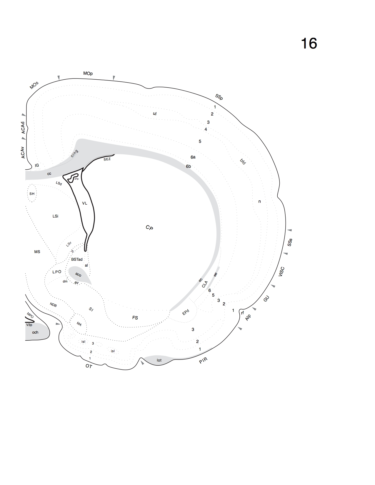
The atlas establishes a foundation for comparing the brain organization of two widely used laboratory mouse substrains. With the rise of transgenic models and the need to benchmark structural differences across hundreds of inbred lines, researchers required a consistent stereotaxic reference. The Mouse Brains Atlas supplies 49 cytoarchitecture and 6 myeloarchitecture levels for each strain, complete with alphabetical indices and comparative lists that map structures by name and bregma level.
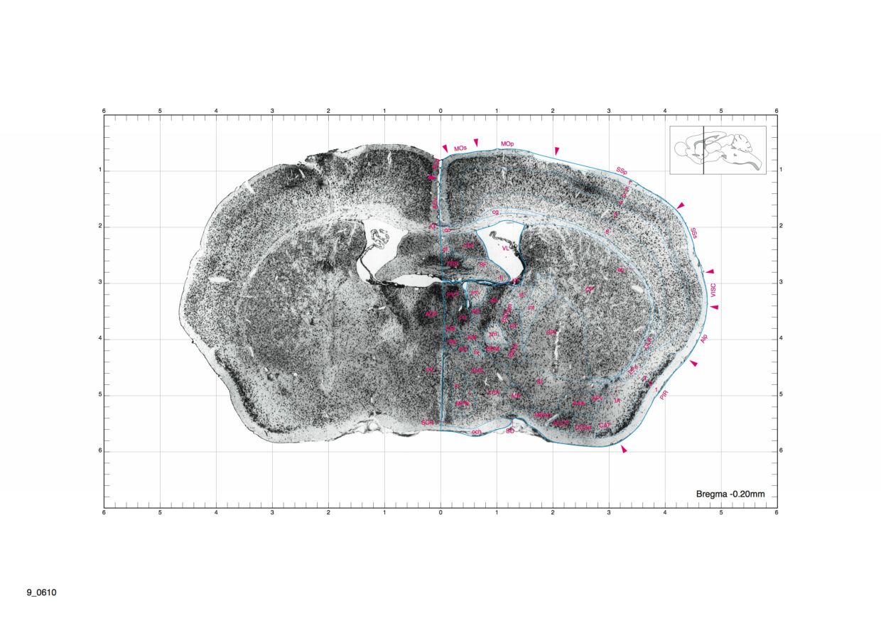
Each plate captures nuclei, cortical areas, and fiber tracts, enabling rapid cross-referencing between strains. Matching CD-ROMs supplement the printed material with Adobe Illustrator files, structure lists, and comparative indices so laboratories can integrate the data directly into their workflows.
Software
The companion application runs on both macOS and Windows, allowing teams to open data files, search structures, and visualize annotated regions.

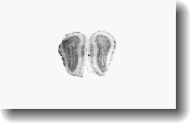
A sample section from level 16 of the coronal series.
A stained section showing neuronal outlines captured for comparison.
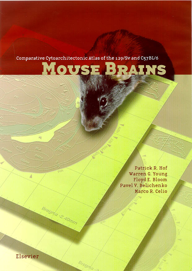
Cover art prepared for the printed atlas.
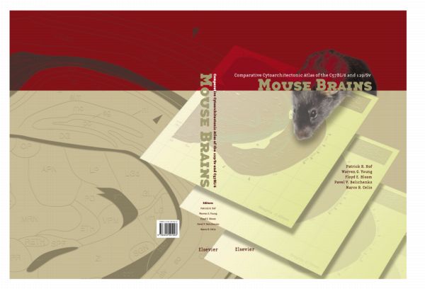
Alternate cover exploration highlighting comparative sections.
(0:04) Animated sweep through coronal sections.
Documentation
Access the final User Manual distributed with the atlas and companion software.